The Figs. show young cells from the threshold epithelium of Utricularia sandersonii. The cell divisions (mitosis) leading to the epithelium’s three-celled glandular hairs begin at the edges of the epithelium and proceed towards the middle region. When the outermost glands are mature, the differentiation stop for the trichomes in the middle zone. In the picture above counted from left the following is seen, a two-celled immature trichome from the velum zone, a newly divided mother cell, where the new cell wall is not yet completed, two mother cells from the transition zone, and two two-celled immature trichomes from the slime producing inner zone. N, nucleus. m, mitochondrion. P, plastid. V, vacuole.
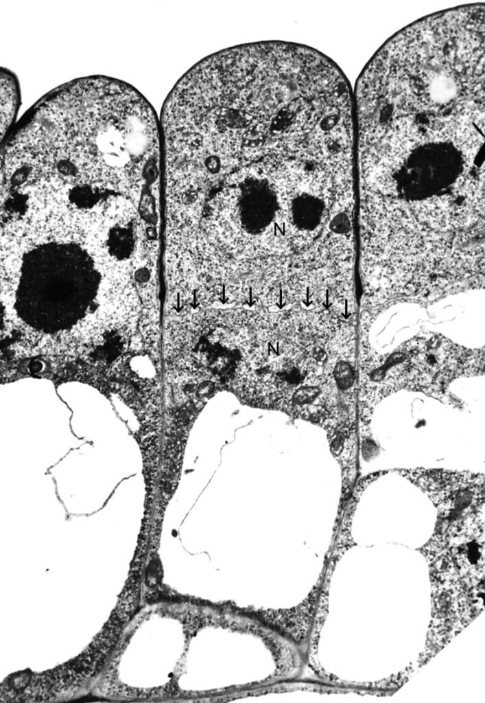
The Fig. to the left shows a newly divided mother cell from the inner part of the threshold epithelium. The new cell-wall (arrows) is not yet finished. Notice that the new wall is positioned at the same level as in the cell to the right. The lower one of the newly divided cell becomes the stalk cell of a glandular trichome and the upper cell becomes the glandular head cell.