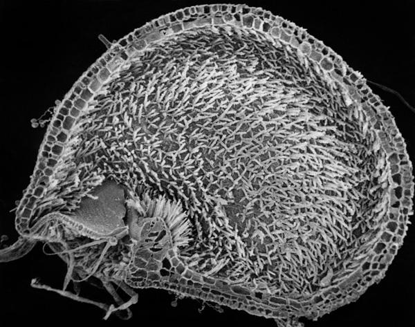
Utricularia australis, longitudinal section of bladder with numerous 4-forked trichomes.
The bladder lumen of Utricularia is equipped with two kinds of glandular hairs functioning partly as enzyme secreting glands partly as absorbing glands and therefore they are called digestive glands. These glands (trichomes) are built over the usual three-part plan with a basal cell, an endodermoid stalk cell (a cell with cutinized lateral walls), and a glandular head consisting of two or four cells. The trichomes are called two- and four-armed hairs. There are far most of the four-armed hairs, which cover the inner surface of the bladder except the door and the threshold. In most species, the two-armed hairs are solely placed on the inside of the threshold.
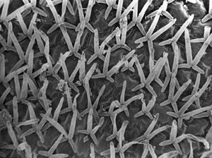
U. australis, 4-forked digestive glands.
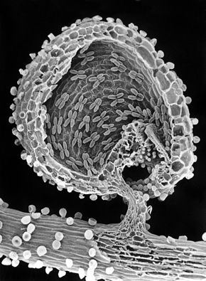
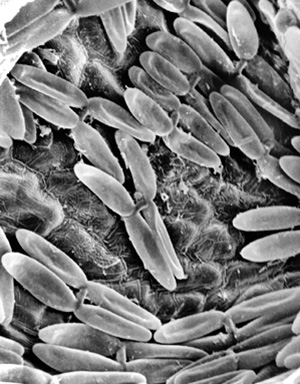
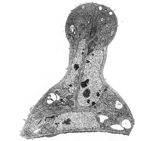
Utricularia sandersonii, bladder lumen with 4-forked digestive glands.
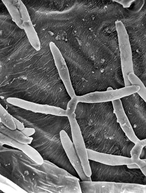
Utricularia reniformis, bladder lumen with 4-forked digestive glands.
The digestive glands secrete the following enzymes: esterase, acid phosphatase, and protease but in addition caught bacteria contribute with other enzymes. In Utricularia vulgaris (Greater Bladderwort) it takes about 10 days to digest a small mosquito larva. After the digestion, the bladder will wilt but with smaller prey, the bladder can be reused several times.
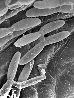
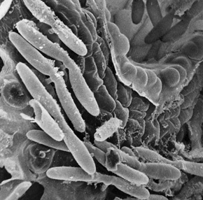
There are considerable variation in the closeness of the digestive glands and in the shape, length, and angel with one another and with the bladder wall. According to taxonomists, these differences alone are sufficient to identify all 225 species. The first nine photos on this page illustrate these differences Utricularia australis, U. sandersonii, U. reniformis, U. subulata, and U. alpine.
Utricularia alpina, bladder lumen with 4-forked digestive glands.
U. sandersonii, 4-forked digestive glands.
U. sandersonii, 2-forked digestive glands.
U. reniformis, 4-forked digestive glands.
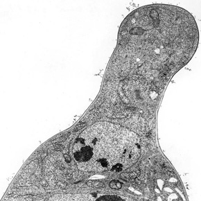
U. australis, 2-forked digestive glands.
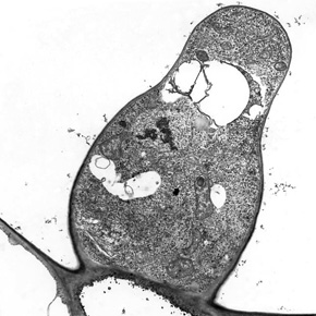
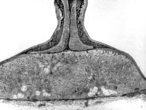
Both types of digestive glands have a remarkable construction, since the arm-cells (glandular cells) are equipped with a neck constriction near the base and a foot below. The foot rests on the endodermoid stalk cell’s very well developed labyrinth wall (diagram and two photos below). Labyrinth walls characterize cells with intensive transport across the cell membrane. The arm cells are thought to both secrete enzymes and absorb nutrients but their labyrinth walls are of modest dimension. However, if the number of four-armed hairs and the total length of the arms are taken into consideration, the area of their cell membrane corresponds so abundantly the membrane area of the stalk cells.
The function of the neck constriction is unknown but two purposes should be considered. When water is pumped out of the bladder after a catch, the enzymes become concentrated in the reduced volume of the bladder and digestion becomes more effective. Secondly, at the beginning the prey will fight to escape and thereby bump into the two- and four-armed glands. The neck constriction is assumed to make the arm-cells more flexible so they give in to the mechanical bumps. If not so, the bladder wall may be slightly deformed with the danger that the door is activated uselessly and water sucked in to dilute the enzyme solution, and more energy must be used to reestablish the vacuum.
U. alpina, 4-forked digestive glands.
U. australis, diagram of 4- and 2-forked trichomes.
It is unknown, if the enzymes are secreted continuously or only if prey is present in the trap as is the case in Pinguicula (Butterwort) and several other carnivorous plants. If a stimulation is required, a vast of enzymes could be prevented when the door opens in vain. A mechanical stimulation system will be of some advantage in relation to a chemical system. A chemical stimulator may e.g. probably not reach a concentration corresponding to the direct secretion on the prey from the digestive glands of Pinguicula. The assumed ability of the arm cells to move when prey bumps into them could be part of a stimulation system. This could either be through action potentials as in the Sundew family (Droseraceae) or through pressure impact on the endoplasmic reticulum in the area of the neck constriction followed by hormone regulated initiation of an ion pump (known from root cap cells) that could flush out enzymes to the bladder lumen.
Utricularia sandersonii, mothercell of a 2-forked glandular trichome.
U. sandersonii, the first cell divison of the mothercell has taken place. the neck constiction is visible.
Left and right show three stages in the development of a digestive gland. It is unknown if there is a division of labor between the two-armed and four-armed glands. One theory says that the two-armed hairs, which only are present on the inside of the threshold, collaborate with the hydathodes in the vestibule and that most of the water is pumped out of the bladder through these glands.
U. sandersonii, 4-forked trichome with stalk cell and two cells in the glandular head. Three cell nuclei are visible.
Utricularia australis, 2-forked digestive gland. Stalk cell with nucleus and labyrinth cell wall. Notice the thick ‘spongy’ wall at the neck constriction.
Utricularia australis, stalk cell of 2-forked digestive trichome and below the basal cell. M, mitochondria. P, plasmodesm. V, labyrint wall.