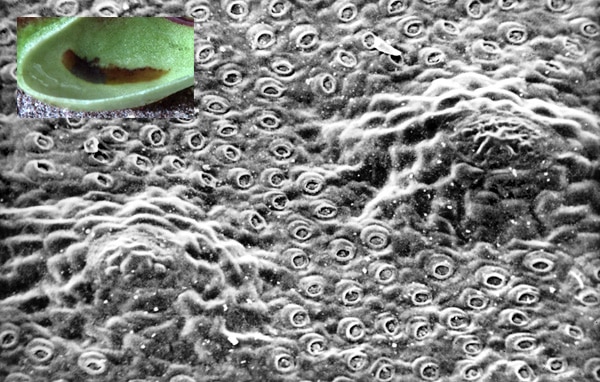
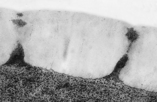
Kirtelvalk på kandens inderside med to store 'vulkankirtler' og talrige små 'stoma-kirtler'.
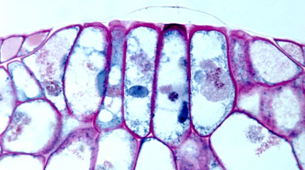
Kirtelvalk med bakterier på epidermiscellers ydervæg, hvor halvdelen er kutiniseret
The brownish gland patches in Cephalotus pitchers are equipped with two types of glands, both assumed to be digestive glands. The relatively few but large glands consist of around 200 cells and are raised up as low cones and may be called vulcan-glands. The small ones are very numerous and established in connection with a stoma converted guard cells (see here). The gland patches are normally covered by the pitcher fluid and sprinkled with bacteria (photo above), which contributes to the digestion with bacteria.
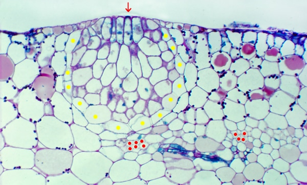
Cross section of ’vulcan-gland’. Yellow dot, endodermoid cell. Red dot, xylem cell in vascular bundle.
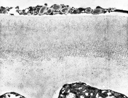
Section of left photo showing cuticle lifted above gland cells. Stained with PAS-aniline-blue-black.
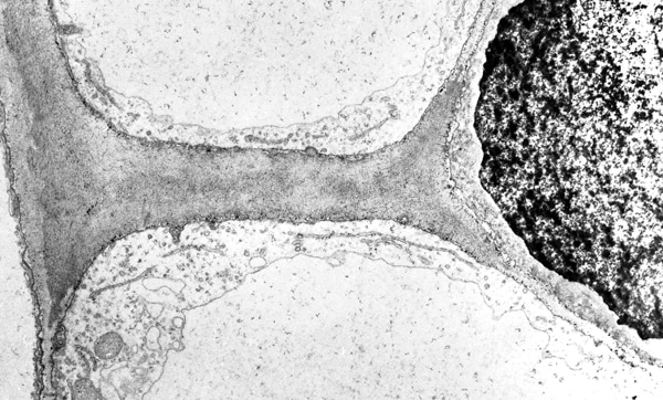
Both gland types are assumed to be digestive glands, but it has not been proven that they function as such. In most carnivorous plants, the digestive glands both secrete digestive enzymes and absorb nutrients. It is noteworthy that in Cephalotus digestive glands are only present on the two gland patches since the patches may not always be covered by pitcher fluid and the bottom zone below the patches are completely free of glands. It has not been investigated if the bottom zone, where the nutrients are concentrated, functions as one large absorptive gland as in the Sarracenia family (see here). According to their structure, the large vulcan-glands seem primarily to be secreting glands. Protease and acid phosphatase are detected in young closed pitchers and these enzymes may originate from the vulcan-glands. These glands function as described for the nectaries on the peristome teeth. The gland cells are incapsulated by endodermoid cells where the radial cell walls on the color-photo at left appear translucent due to all interfibrillar space being blocked by the lipid cutin. Vascular bundles come as close as one cell layer from the endodermoid cells. Phloem is seen between the two marked vascular bundles. The photo below at left shows the cuticle on the surface of the central glandular cells is penetrated by channels consisting of cellulose and pectin, which allows the passage of enzymes. Furthermore, the pressure below the cuticle may be so large that the cuticle is lifted up from the cell wall as a balloon (red arrow on left photo). This opens the channels further.
The cuticle on central glandcell is penetrated by cellulose-pectin channels.
Cutinized cell wall between two endodermoid cells. Gland cell at right.
H. S. Heide-Jørgensen, april 2014, translated May 2021.