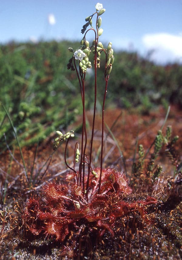
Rundbladet soldug er den mest udbredte soldug art i Danmark. Den er cirkumpolar inklusive det sydlige Grønland. Den trives fint med konkurrence fra tørvemos (Sphagnum). På billedet th. vokser soldug tæt rundt om en grundvands regulere klitlavning i den rødbrune bræmme, hvor der hverken er for vådt eller tørt.
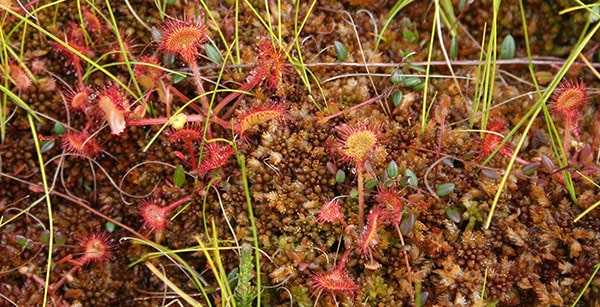
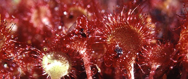
Drosera rotundifolia blomstrer i august. De 5-tallige blomster er kun åbne midt på dagen, og de sidder højt hævet over fælderne. I tørt og solrigt vejr kan slimen på tentaklerne tørre ud. På billedet herunder ses i midten et blad foldet sammen om et bytte som resultat af den på hovedsiden omtalte auxin regulerede vækstbevægelse.
Langbladet soldug - Drosera anglica
Langbladet soldug har i Danmark kun få voksesteder på højmoser og i fugtige klitheder i Jylland. Dens totale udbredelse er næsten cirkumpolar. Den findes dog ikke i Island og Grønland, men i Nordamerika når den op i Alaska. Der findes også nogle subtropiske forekomster på Hawaii og i det sydlige Europa og sydlige Japan. Den trives fint i sure omgivelser mellem Sphagnum. Den overvintrer ved vinterknopper kaldet hibernakler. Arten er selvbestøende og menes opstået som en diploid hybrid mellem den nordamerikanske D. linearis og D. rotundifolia. Drosera anglica x obovata er en steril hybrid med D. rotundifolia. Hybriden forveksles undertiden med Drosera intermedia.
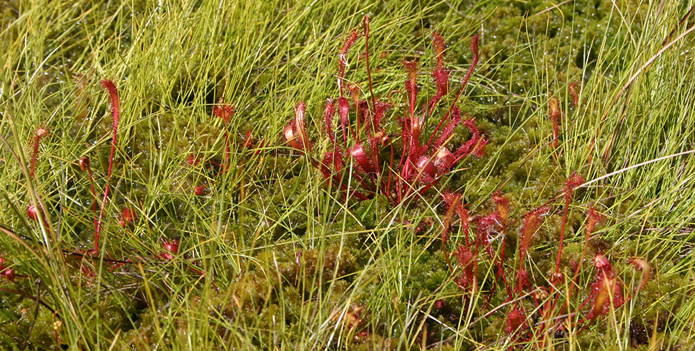
Liden soldug - Drosera intermedia
Liden soldug findes især i moser, hvor den står under vådere forhold end de to øvrige danske arter, rundbladet og langbladet soldug. Den har hovedudbredelse i det vestlige Europa og østlige Nordamerika, men når ned i det nordlige Sydamerika. I tempereret klima overvintrer den ved hibernakler. Bladene er oprette, og formen er intermediær mellem de to førnævnte arter. Blomsterstanden virker sidestillet, idet den er bøjet til siden, mens den står opret centralt i rosetten hos de to andre danske arter. Bomstrer fra juni til august. På billedet t.h. vokser den sammen med blærerod Utricularia australis. – Drosera anglica x obovata er en steril hybrid med D. rotundifolia. Hybriden forveksles undertiden med Drosera intermedia.
Heide-Jørgensen H S, Kuijt J. 1993. Epidermal derivatives as xylem elements and transfer cells: a study of the host-parasite interface of two species of Triphysaria (Scrophulariaceae). - Protoplasma 174: 173-183.
Abstract/Summary: Haustoria of Triphysaria pusilla and T. versicolor subsp. faucibarbata from a natural habitat were analysed by light and electron microscopy. The keel-shaped edge of the secondary haustorium generally splits the epidermis and cortex of the host root parallel to the root axis, and penetrates to the host vascular tissue. Anticlinally elongated epidermal cells of the haustorium constitute most of the host/parasite interface. Some of these epidermal cells are divided by oblique cell walls. Some of their oblique daughter cells as well as some undivided epidermal cells differentiate into xylem elements. Single epidermal cells occasionally intrude into the vascular tissue of the host and individual host cells can be invaded. The surface area of the plasmalemma in parasitic parenchymatous interface cells is increased by the differentiation of wall labyrinths characteristic of transfer cells and by the development of membrane-lined cytoplasmic tubules or flattened sacs which become embedded in the partly lignified interface cell-wall. Mycorrhizal fungal hyphae enter the xylem bridge in some haustoria. Implications of these observations for the function of the haustorium are discussed.
Transmissionelectron micrograph of mature haustorium in transverse section of parasite and host root at the haustorial axis. – Glutaraldehyde/osmium fixed material contrasted with uranyl acetate and lead citrate.
Thick arrows, oblique cell walls between daughter cells of one epidermal mother cell.
Thin arrows, electron-dense material lining secondary wall thickenings.
F, fungal cell.
IP, parasite interface parenchyma cell originating from epidermal cell layer.
LW, lignified secondary cell wall in xylem element.
Bar, 10 mµ
XW, xylem bridge vessel member.
FIGUR LEGEND abs 19:
Transmissionelectron micrograph of mature haustorium in transverse section of parasite and host root at the haustorial axis. – Glutaraldehyde/osmium fixed material contrasted with uranyl acetate and lead citrate.
Thick arrows, oblique cell walls between daughter cells of one epidermal mother cell.
Thin arrows, electron-dense material lining secondary wall thickenings.
F, fungal cell.
IP, parasite interface parenchyma cell originating from epidermal cell layer.
LW, lignified secondary cell wall in xylem element.
Bar, 10 mµ
XW, xylem bridge vessel member.
Rettelser/Corrigenda: Missing abbreviation in Figure legends page 174: L, lignified secondary cell wall in xylem element.