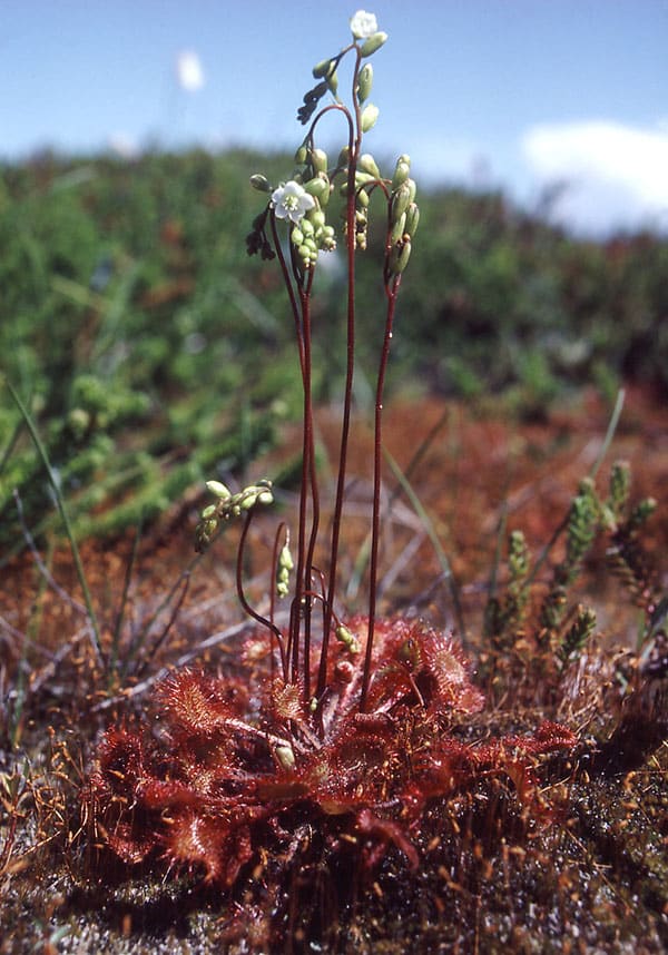
Rundbladet soldug er den mest udbredte soldug art i Danmark. Den er cirkumpolar inklusive det sydlige Grønland. Den trives fint med konkurrence fra tørvemos (Sphagnum). På billedet th. vokser soldug tæt rundt om en grundvands regulere klitlavning i den rødbrune bræmme, hvor der hverken er for vådt eller tørt.
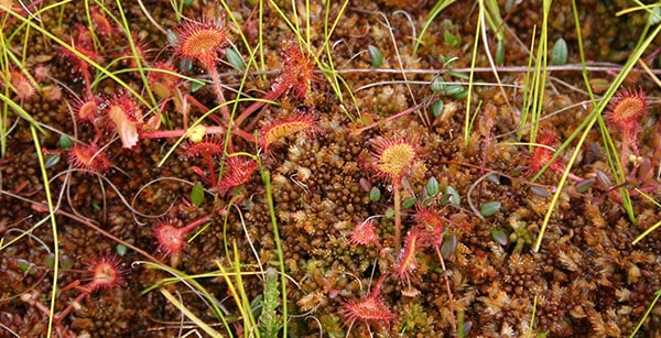
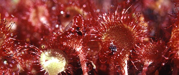
Drosera rotundifolia blomstrer i august. De 5-tallige blomster er kun åbne midt på dagen, og de sidder højt hævet over fælderne. I tørt og solrigt vejr kan slimen på tentaklerne tørre ud. På billedet herunder ses i midten et blad foldet sammen om et bytte som resultat af den på hovedsiden omtalte auxin regulerede vækstbevægelse.
Langbladet soldug - Drosera anglica
Langbladet soldug har i Danmark kun få voksesteder på højmoser og i fugtige klitheder i Jylland. Dens totale udbredelse er næsten cirkumpolar. Den findes dog ikke i Island og Grønland, men i Nordamerika når den op i Alaska. Der findes også nogle subtropiske forekomster på Hawaii og i det sydlige Europa og sydlige Japan. Den trives fint i sure omgivelser mellem Sphagnum. Den overvintrer ved vinterknopper kaldet hibernakler. Arten er selvbestøende og menes opstået som en diploid hybrid mellem den nordamerikanske D. linearis og D. rotundifolia. Drosera anglica x obovata er en steril hybrid med D. rotundifolia. Hybriden forveksles undertiden med Drosera intermedia.
Liden soldug - Drosera intermedia
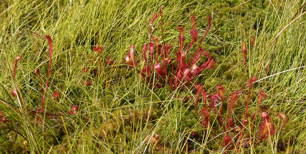
Liden soldug findes især i moser, hvor den står under vådere forhold end de to øvrige danske arter, rundbladet og langbladet soldug. Den har hovedudbredelse i det vestlige Europa og østlige Nordamerika, men når ned i det nordlige Sydamerika. I tempereret klima overvintrer den ved hibernakler. Bladene er oprette, og formen er intermediær mellem de to førnævnte arter. Blomsterstanden virker sidestillet, idet den er bøjet til siden, mens den står opret centralt i rosetten hos de to andre danske arter. Bomstrer fra juni til august. På billedet t.h. vokser den sammen med blærerod Utricularia australis. – Drosera anglica x obovata er en steril hybrid med D. rotundifolia. Hybriden forveksles undertiden med Drosera intermedia.
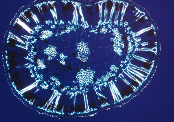
Transverse section of mature leaf with sclereids in the palisade tissue shown in polarized light.
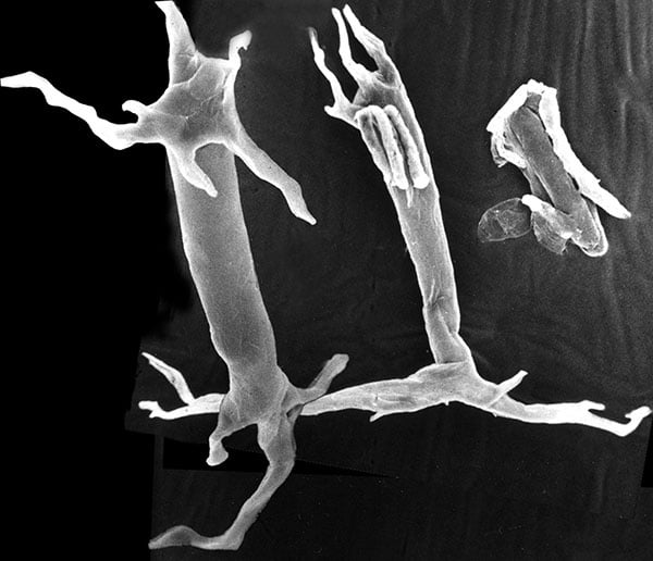
SEM of two sclereids (epidermal end up) from mature leaf macerated in nitric acid and chromic acid.
Artifacts and disturbing structures removed from original photo.
Heide-Jørgensen, H S. 1990. Xeromorphic leaves of Hakea suaveolens R. Br. IV. Ontogeny, structure and function of the sclereids. - Aust. J. Bot. 38: 25-43.
Abstract/Summary: Osteosclereids in the leaves of Hakea suaveolens are investigated from a developmental, structural and functional point of view. The sclereid initial cell is located outermost in a boundary parenchyma comprising 1-3 cell layers next to the palisade tissue. Boundary parenchyma cells, including sclereid initial cells, are the first cells to accumulate starch. Acid phosphatase was localised during development of sclereids and palisade cells. The outer ramifications of the sclereid form a pseudohypodermis, and the inner ramifications penetrate 1-3 cell layers inwards, making contact with specialised cells (tracheoids) of the vein endings and with fibres. The sclereids do not become lignified in the outer ramifications, and they remain alive as long as the leaf itself. Water pathways were localised by addition of the fluorochrome berberine sulfate to the transpiration stream. It is concluded that, besides giving mechanical strength, the sclereids are also important as vein extensions and that, like bundle sheath extensions, they conduct water to the epidermis and directly to both palisade layers. The percentage of the cross-sectional leaf area occupied by lignified water-conducting cells, including sclereids, is higher than in many other cylindrical, xeromorphic leaves. There are about 200 sclereids per square millimetre of palisade tissue in sun leaves, but the number falls in shade leaves and after fertilisation with nitrate and phosphate.
Rettelser/Corrigenda: According to Flora of Australia Hakea suaveolens R. Br. is now named Hakea drupacea (C. F. Gaertn.) Roem. & Schult.